Pulmonary Embolism (PE) - Medical Notes

By Dr Nehana Gittens (FY2)
Pulmonary Embolism (PE)
A venous thrombus embolism (VTE) is a blood clot that forms in the venous circulation.
The clot can then disrupt blood flow which can become life-threatening and potentially fatal. The types of VTE include deep vein thrombosis (DVT) and a pulmonary embolism (PE). This article will focus on PE.
PE may occur in the absence of DVT (20-30%)
Click here to read our article on Deep Vein Thrombosis (DVT)
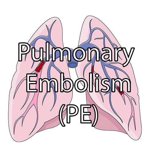
Pulmonary embolism occurs as a result of a blood clot forming in the deep veins which then embolises (breaks off) and becomes lodged in the pulmonary arteries. This can be a life-threatening event. The blood clot can block pulmonary circulation and put strain on the right side of the heart.
- Shortness of breath
- Chest pain (pleuritic)
- Cough
- Haemoptysis
- Hypoxia
- Haemodynamic instability
- Tachycardia
- Hypotension
- Acute right heart strain -> raised JVP
- Syncope
Virchow’s Triad
Virchow’s Triad (as mentioned in the DVT article) explains the factors that increase the risk of a blood clot forming. The factors include: hypercoagulability of blood, stasis of blood flow and injury to vessel wall. The effect of these factors on blood flow can increase blood coagulation. Risk factors for clotting fall under these major factors for clotting, such as immobility which causes stasis of blood flow.
Risk Factors
Key risk factors for a pulmonary embolism include:
- Immobility
- Recent surgery
- Long haul travel
- Malignancy
- Pregnancy
- Thrombophilia e.g antiphospholipid syndrome, Factor V Leiden
- Hormone estrogen therapy
- Trauma
Investigations
When a PE is clinically suspected a Wells Score for suspected PE is calculated. This gives an indication as to whether a PE is likely or not.
For a detailed breakdown of the Wells criteria and to use the score calculator, click here
D dimer
- This is a blood test for protein in blood when clots dissolve
- It is sensitive but not specific; can be raised in other conditions e.g infection
- Rules in an PE but does not confirm it
CT Pulmonary Angiogram
Identifies the clot in pulmonary circulation and is the definitive investigation.
Chest X-Ray
Used to rule out other pathology e.g pneumonia.
V/Q Scan
Can be used in pregnancy and with reduced renal function.
Management
PEs are treated with anticoagulation. Treatment dose anticoagulation is recommended to be started immediately when VTE is suspected. DOACs are more commonly used first line such as apixaban or rivaroxaban. Alternatively, LMWH (low molecular weight heparins) can be used e.g enoxaparin.
Dosing Example of Apixaban for PE
10mg BD for 7 days, then 5mg BD till the end of therapy
Massive PE
- Treated with continuous infusion of unfractionated heparin
- Thrombolysis is also used to break down the clot
Length of Treatment
- 3 months for patients with identifiable/reversible cause of VTE
- 3-6 months for patients with active cancer
- Long-term for patients with unprovoked VTE, recurrent VTE or an irreversible underlying cause e.g thrombophilia
References
- BMJ Best Practice. (2024). VTE treatment algorithm. Available at: https://bestpractice.bmj.com/topics/en-gb/3000201/treatment-algorithm (Accessed: 18 February 2024).
- NICE. (2020). Oral anticoagulation prescribing in DVT and PE. Available at: https://cks.nice.org.uk/topics/deep-vein-thrombosis/prescribing-information/oral-anticoagulants/ (Accessed: 21 February 2024).
Image credits
PE illustration (cover) by Laboratoires Servier, CC BY-SA 3.0 <https://creativecommons.org/licenses/by-sa/3.0>, via Wikimedia Commons


Leave a comment